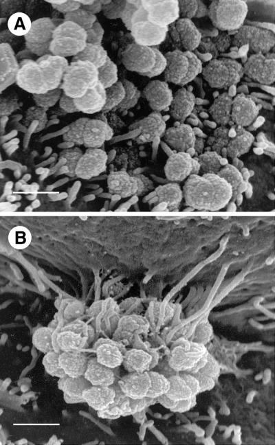
FIG. 3.
HEC-1-B cell microvilli adhere to P+ Opa+ gonococci along the full lengths of the microvilli. (A) At this SEM magnification it is clear that after 6 h of incubation only the distal ends of microvilli adhere to MS11mk P− Opa+ organisms. Microvilli have not been elongated. Both true diplococci and adherent individual cocci are visible. (B) In this SEM photomicrograph microvilli can be seen to have wrapped themselves tightly around MS11mk P+ Opa+ cocci in the microcolony after 6 h of incubation on HEC-1-B cells. Microvilli adhere tightly to individual cocci up to the full lengths of the microvilli. Microvillus adherence is clearly separate from bacterium-pilus interactions. Microvilli at the top of the micrograph appear to have pulled the cell surface up along the side of the colony, creating the appearance of a microcolony hanging from a pedestal of microvilli. (The electron beam is focused vertically, not horizontally.) The extent of microvillus elongation is remarkable, and the contrast between microvillar responses to piliated and nonpiliated Opa+ bacteria is striking. Magnification, ×15,500; bar, 1 μm.

