Figure 5.

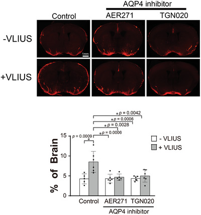
The role of AQP4 in VLIUS‐induced glymphatic circulation. Fluorescence imaging and quantification of the area covered by the tracer influx in coronal brain slices revealed that 30 min after intracisternal injection, paravascular cerebrospinal fluid influx increased with very low‐intensity ultrasound (VLIUS) stimulation, and did not increase with the co‐administration of an aquaporin‐4 (AQP4) inhibitor (AER271 and TGN020). Dots represent individual mice in each group (control group, n = 5; VLIUS group, n = 6; AQP4 inhibitor group, n = 6; and AQP4 inhibitor + VLIUS group, n = 5). Scale bar: 1 mm. The results, for which the data are presented as the mean ± SD (error bars denote SD), were analyzed using ANOVA followed by Tukey's post‐hoc test to assess between‐group differences. An asterisk indicates p < 0.05.
