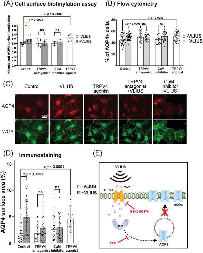
Figure 7.

AQP4 protein translocation to the cell surface increased after VLIUS stimulation. A) The mean fold change in aquaporin‐4 (AQP4) surface expression, measured by cell‐surface biotinylation in C6 cells. The calmodulin (CaM) inhibitor was 20.8 µM trifluoperazine (TFP). The transient receptor potential vanilloid‐4 (TRPV4) antagonist was 100 nM GSK2193874, while the TRPV4 agonist was 2 µM GSK1016790A. Cells had been pre‐incubated with drug 30 min before very low‐intensity ultrasound (VLIUS) treatment. AQP4 on the cell surface significantly increases after treatment with VLIUS or TRPV4 agonist. However, administration of TRPV4 antagonist or CaM inhibitor inhibits VLIUS‐facilitated AQP4 cell surface localization. B) The AQP4 surface expression is analyzed by flow cytometry (without permeabilization). The population of AQP4‐positive cells was significantly increased by VLIUS or TRPV4 agonists. Administration of TRPV4 antagonist or CaM inhibitor inhibits the VLIUS effect. C) AQP4 on the cell surface can also be observed by immunofluorescence staining (without permeabilization). Cells had been counterstained by WGA‐conjugated Alexa Fluor™ 488 to determine cell boundaries. Scale bar: 10 µm. D) Quantification of AQP4 on the cell surface using immunofluorescence staining. E) The schematic showing that both the CaM inhibitor and the TRPV4 antagonist can inhibit VLIUS‐induced AQP4 translocation to the cell surface. The results, for which the data are presented as the mean ± SD (error bars denote SD), shown in Figure 7 (A,B,D) were analyzed using Kruskal‐Wallis test followed by Dunn's post‐hoc test to assess between‐group differences. An asterisk indicates p < 0.05.
