Fig. 2.

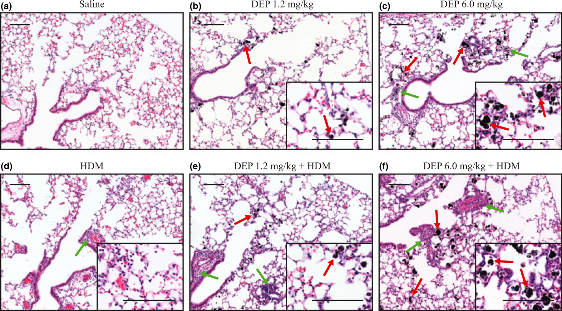
Lung histology following DEP and allergen exposure. (a–f) Haematoxylin and eosin staining of lung sections showing DEP (red arrows) and inflammation (green arrows) in the small airways and alveolar regions of the lung; scale bar = 100 μm.

Lung histology following DEP and allergen exposure. (a–f) Haematoxylin and eosin staining of lung sections showing DEP (red arrows) and inflammation (green arrows) in the small airways and alveolar regions of the lung; scale bar = 100 μm.