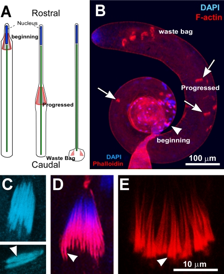
Figure 1.
The stages of sperm individualization in wild-type testis. (A) Schematic illustrates relative positions of F-actin cones (marked in red) within a single spermatid cell at different stages of individualization. (B) RITC:phalloidin (red) and DAPI (blue) staining revealed matured nuclear bundles and the beginning stage ICs (arrowhead) at the basal region, progressed ICs (arrows) along the length of the testis and the waste bags near the apical tip. (C) Nuclear bundle (upper panel) and isolated nuclei (arrowhead) from a postelongation stage cyst. (D) Nuclear bundle and freshly formed investment cones (arrowheads) at the early individualization stage. (E) Bundle of progressed investment cones (arrowhead). C, D and E are presented at the same scale as indicated in E.

