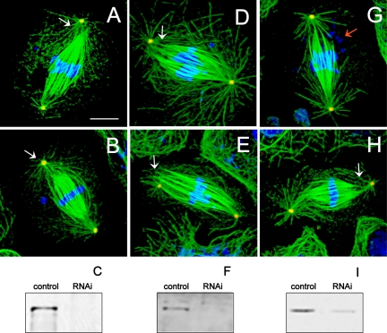
Figure 2.
Defects in spindle pole organization in dynein–dynactin-depleted cells. Immunofluorescence localization of α-tubulin (green), γ-tubulin (red), and DNA (blue). Cells depleted of DHC (A and B), DIC (D and E), and p50 dynamitin (G and H) displayed longer than normal metaphase spindles with unfocused microtubules at spindle poles and loosely attached or completely detached centrosomes (white arrows). Some spindles displayed defects in chromosome alignment at the metaphase plate and apparent chromosome fragmentation (red arrow). Bar, 10 μm. Immunoblots of S2 cell cultures probed with anti-DHC (C), anti-DIC (F), and anti-p50 dynamitin (I) antibodies show 97, 82, and 80% depletion of protein, respectively (confirmed by densitometry) after dsRNA treatment. All lanes loaded with 50 μg of protein.

