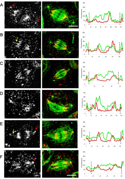
Figure 9.
Localization and linescan quantitation of Asp in dynein-depleted S2 cells. After DHC depletion by using RNAi (Figure 2), Asp localization was significantly perturbed. We saw a marked accumulation at the centrosomes (A, E, and F white-gray panels; red arrows) “wispy” fibers of Asp connecting centrosomes to half-spindles (B and E; yellow arrows) and a more diffuse localization of Asp throughout the spindles, including the central spindle (D and F). Bars, 10 μm. Corresponding line scans of fluorescence intensity (tubulin, green; Asp, red) along the pole-to-pole axis of spindles from A to F clearly reveal a loss of Asp from the transition zones between centrosomes and spindles poles, with a corresponding accumulation of the protein at the centrosomes (black dotted lines).

