Figure 7.
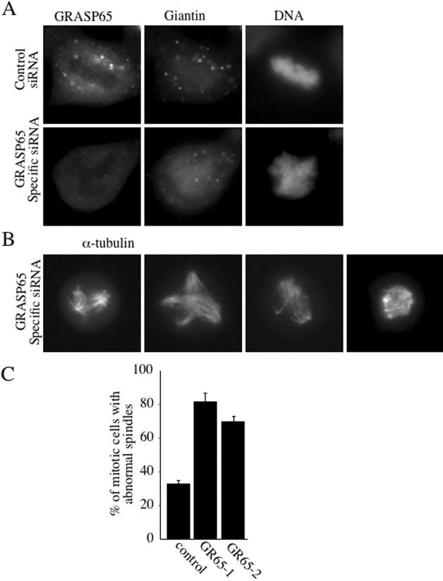
GRASP65 depletion promotes spindle abnormalities. (A) A nonsynchronous population of cells was processed for immunofluorescence microscopy 48 h after transfection with scrambled control or GRASP65-specific siRNA, using antibodies to GRASP65 to verify the knockdown, giantin to monitor the organization of Golgi membranes, and with the DNA dye Hoechst to identify mitotic cells with condensed chromatin. (B) Mitotic GRASP65-depleted HeLa cells were stained with anti-α-tubulin antibody to reveal the organization of the mitotic spindle. Four different mitotic cells are shown. (C) The percentage of mitotic cells with abnormal spindles (monopolar, multipolar, misaligned, unfocused pole) observed 48 h after transfection with GR65–1–scrambled, GR65–1 or GR65–2. These results are derived from three independent experiments, each sample derived from two independent transfections.

