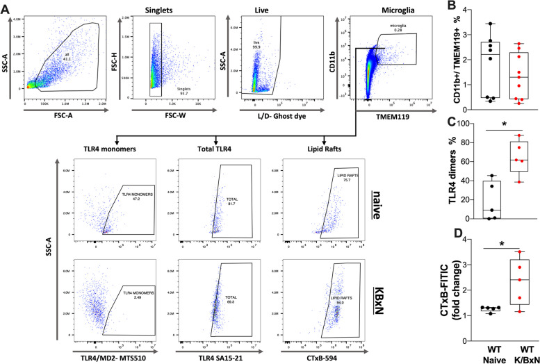
Fig. 9.
Microglia from arthritic mice show increased TLR4 dimers and lipid rafts. Single cell suspensions from spinal cords were generated from naïve and WT K/BxN male mice injected with serum after 28 days and stained for microglia markers, TLR4 and lipid rafts. (A) Gating strategy and representative plots of microglia (CD11b + /TMEM119 +) from K/BXN serum injected and naïve mice showing cell population intensity (mean fluorescence intensity), for TLR4 monomers (MTS510 clone), total TLR4 (SA15-21 clone) and lipid rafts (CTxB binding). Quantification of microglia populations (B), TLR4 dimers (C) and change in lipid rafts (D) are shown (n = 5–9 per group). There were no significant changes in the numbers of CD11b + /TMEM119 + cells at 28 days; however, the percentage of TLR4 dimers (p < 0.015) and increase in the lipid rafts were significant (p = 0.033, Students t test)

