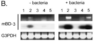
FIG. 3.

Analysis of mBD-3 expression by dot blot analysis and RT-PCR. (A) Measurement of mBD-3 expression by dot blot analysis. A filter dotted with mRNA from a number of mouse tissues was hybridized to an mBD-3 probe. Signals were quantified with a PhosphorImager system and normalized to expression of the housekeeping gene ubiquitin. Data are expressed as relative hybridization signals. Only those tissues which demonstrated a significant signal over background are presented, except for skeletal muscle, which is an example of an organ with low signal. The experiment was repeated on three occasions with virtually identical results. (B) Detection of mBD-3 expression was measured before and after intratracheal injection of P. aeruginosa PAO1 in various mouse tissues by RT-PCR. Poly(A)+ RNA was isolated from mouse tissues and reverse transcribed, and the cDNAs were amplified by using mBD-3-specific primers. A single 270-bp band visualized directly by staining with ethidium bromide was generated by the amplification (upper panel). The PCR products were blotted onto nitrocellulose filter and hybridized with radiolabeled mBD-3 cDNA (middle panel). The mRNA coding for G3PDH was amplified by using gene-specific primers (lower panel). Lanes: 1, heart; 2, lung/trachea; 3, kidney; 4, small bowel; and 5, liver.

