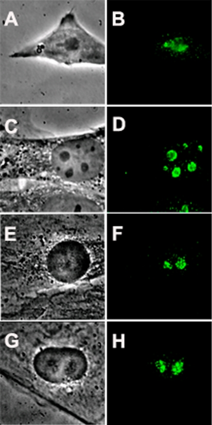
Figure 1.
Nucleolar localization of nucleostemin. Murine and rat cells were stained with a peptide antibody raised against mouse nucleostemin (Tsai and McKay, 2002) followed by detection of the primary antibody with fluorescein-conjugated anti-chicken IgY. A, C, E, and G are phase contrast micrographs, and B, D, F, and H are the corresponding immunofluorescence images. (A and B) Murine embryonic stem cells. (C and D) Mouse 3T3 cells. (E and F) Rat L6 myoblasts. (G and H) Rat NRK cells. Each panel shows a microscope field 35 μm in width.

