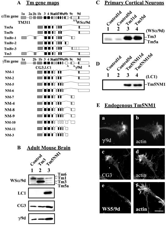
Figure 1.
Organization of Tm genes, their expression in transgenic mice, and subcellular localization in neurons. (A) Schematic representation of the organization of the α, and γ mammalian Tm genes and the respective neuronal isoforms. Exons, shaded boxes, are numbered 1–9; unshaded boxes correspond to 3′ untranslated sequences; and introns are shown as lines. The black shaded exons are common to all genes. The name of the Tm antibody is indicated in bold below the exon where the epitope is found. (B, C, and D) Equal loading (10 μg) of total cellular protein was isolated and electrophoresed on a 12.5% low-bis SDS-PAGE gel. (B) Immunoblots of adult mouse brain. Lane 1, control brain; lane 2, Tm3 transgenic; and lane 3, Tm5NM1 were probed with WSα/9d to detect Tm3, LC1 to detect the exogenous Tm5NM1, CG3 to detect all products from the γTm gene, and γ/9d to detect Tm5NM1 and Tm5NM2. (C) Immunoblot of Tm3 primary cortical neurons. Lane 1, control 1 d old; lane 2, control 5 d; lane 3, Tm3 1 d; and lane 4, Tm3 5 d, probed with the WSα/9d antibody. (D) Immunoblot of Tm5NM1 primary cortical neurons. Lane 1, control 1 d old; lane 2, control 5 d; lane 3, Tm5NM1 1 d; and lane 4, Tm5NM1 5 d, probed with the LC1 antibody. (E) Endogenous Tm5NM1 sorts to the peripheral region of the growth cone unlike Tm5NM2. Control primary cortical neurons cultured for 5 d were double immunofluorescence stained with γ/9d (a), actin (b), CG3 (c), actin (d), WS5/9d (e), and actin (f). Bar, 10 μm.

