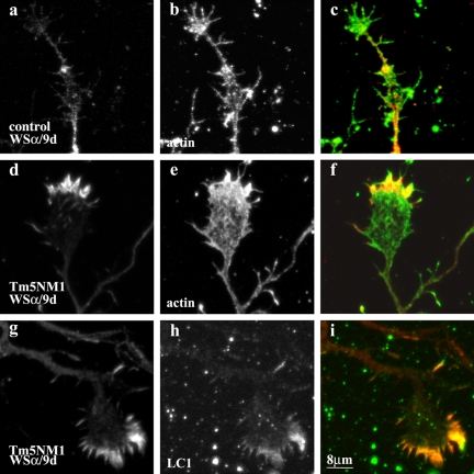
Figure 7.
Recruitment of Tm5a by Tm5NM1 to the growth cone in vitro. Primary cortical neurons were cultured for 5 d and double immunofluorescence stained with WSα/9d that detects Tm5a (a, d, and g) and actin (b and e) or LC1 (h). (c) Merged image of a (red) and b (green). (f) Merged image of d (red) and e (green). (i) Merged image of g (red) and h (green). Note the enrichment of Tm5a in the peripheral region of the Tm5NM1 growth cone (d and g) where actin (e) and the exogenous Tm5NM1 (h) also are enriched. In control neurons, Tm5a (a) is highly diminished in 5-d-old growth cones although actin is present (b). Bar, 8 μm.

