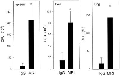
FIG. 3.
M. avium growth in mice treated with anti-CD40L MAb. Mice were infected with 106 M. avium and treated with MR1 (anti-CD40L MAb) or control IgG. M. avium growth in the spleen, liver, and lungs was determined by the CFU assay on day 35. Results shown are mean of CFU per organ ± SD. ∗, P < 0.01 compared to CFU in the organs of M. avium-infected mice treated with control hamster IgG.

