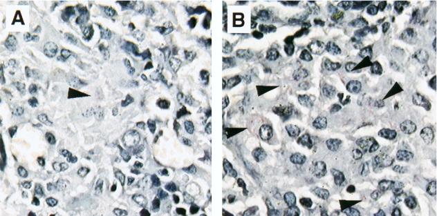
FIG. 4.
Mycobacterial burden in tissue from M. avium-infected mice treated with anti-CD40L MAb. Mice were infected with M. avium and treated with anti-CD40L MAb or control IgG. Mice from both groups were sacrificed on day 35. The spleen was collected from each mouse, sectioned (5 μm), and stained with Ziehl-Neelsen acid-fast stain to visualize mycobacteria. The sections were viewed at a magnification of ×630. Shown are spleen sections from M. avium-infected mice treated with control IgG (A) and anti-CD40L MAb (B). Arrowheads indicate acid-fast bacilli.

