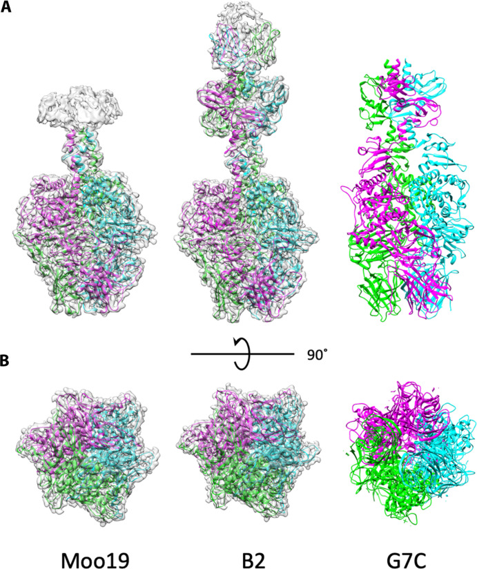
Fig. 5. Structure of isolated tailspikes.
Cryo-EM density maps of the isolated tailspike proteins (gp82 for Moo19 and a complex of gp48/gp49 for B2) are shown with atomic models fitted in. Each structure is a homotrimer, with individual ribbons colored cyan, green, and magenta. (A) Side views. Moo19 and B2 are compared with the tailspike protein in phage G7C [PDB ID: 4QNL (41)]. All three display highly similar topology. (B) 90° rotation showing the distal end of the trimer that interacts with the host cell surface.

