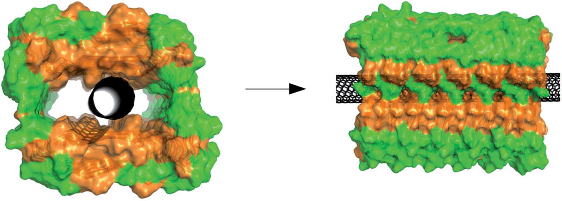
Figure 2.

Illustration of a single walled nano tube (SWNT) located in the hollow core of an amyloid-beta peptide fibril. Orange and green represent hydrophobic and hydrophilic residues, and the black one is the CNT. Although this structure is theoretically possible, it is not backed by experimental evidence
Note. Reprinted from “The Aβ peptide forms non-amyloid fibrils in the presence of carbon nanotubes” by Luo J, Wärmländer SK, Yu CH, Muhammad K, Gräslund A, Pieter Abrahams J. Nanoscale. 2014;6:6720–6 (https://pubs.rsc.org/en/content/articlelanding/2014/nr/c4nr00291a). © 2014 The Royal Society of Chemistry.
