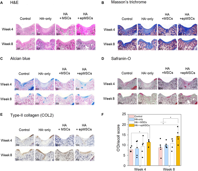
Fig. 6.

Histological and immunohistochemical (IHC) analyses of cartilage repair in the rat model at weeks 4 and 8 after transplantation. (A) Hematoxylin and eosin (H&E), (B) Masson’s trichrome, (C) Alcian blue, and (D) Safranin-O staining images and (E) type II collagen (COL2) IHC staining images of osteochondral defects at weeks 4 and 8 after transplantation. Scale bar = 500 μm. (F) Quantitative data of cartilage regeneration according to the O’Driscoll histological cartilage repair scale with Safranin-O stain at weeks 4 and 8. An asterisk (*) denotes a statistically significant difference (n = 5, *P < 0.05, **P < 0.01).
