FIG. 6.
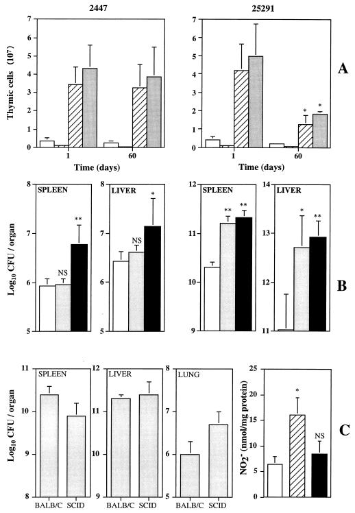
Involvement of the thymus in systemic infection by M. avium 2447 or 25291. (A) Cellularity of the thymus in terms of numbers of CD4+ CD8− (open bars), CD4− CD8+ (solid bars), CD4+ CD8+ (hatched bars), or total cells (shaded bars) of the thymi of mice infected for 1 or 60 days with strains 2447 (left) or 25291 (right). Results are means ± 1 standard deviation (SD). (B) Effects of thymectomy alone (shaded bars) or thymectomy followed by CD4+-T-cell depletion (solid bars) on the proliferation of M. avium 2447 (left) or 25291 (right) compared to that in untreated mice (open bars) at 40 days of infection. The results are shown as means ± 1 SD (n = 5). (C) Infection of SCID mice with M. avium 25291. The growth of M. avium was evaluated in the spleens, livers, and lungs of BALB/c and SCID mice after 30 days of infection. The results are shown as the geometric means ± 1 SD of the CFU values from three or four mice. The degree of macrophage activation of the peritoneal cells from the same animals as evaluated from the production of LPS-stimulated nitrite secretion is shown on the right. In vitro production of NO2− by peritoneal macrophages from uninfected BALB/c mice (open bar), infected BALB/c mice (hatched bar), or infected SCID mice (solid bar) is shown. The data represent means ± 1 SD of triplicates from a pool of three or four mice. ∗, P < 0.05; ∗∗, P < 0.01.

