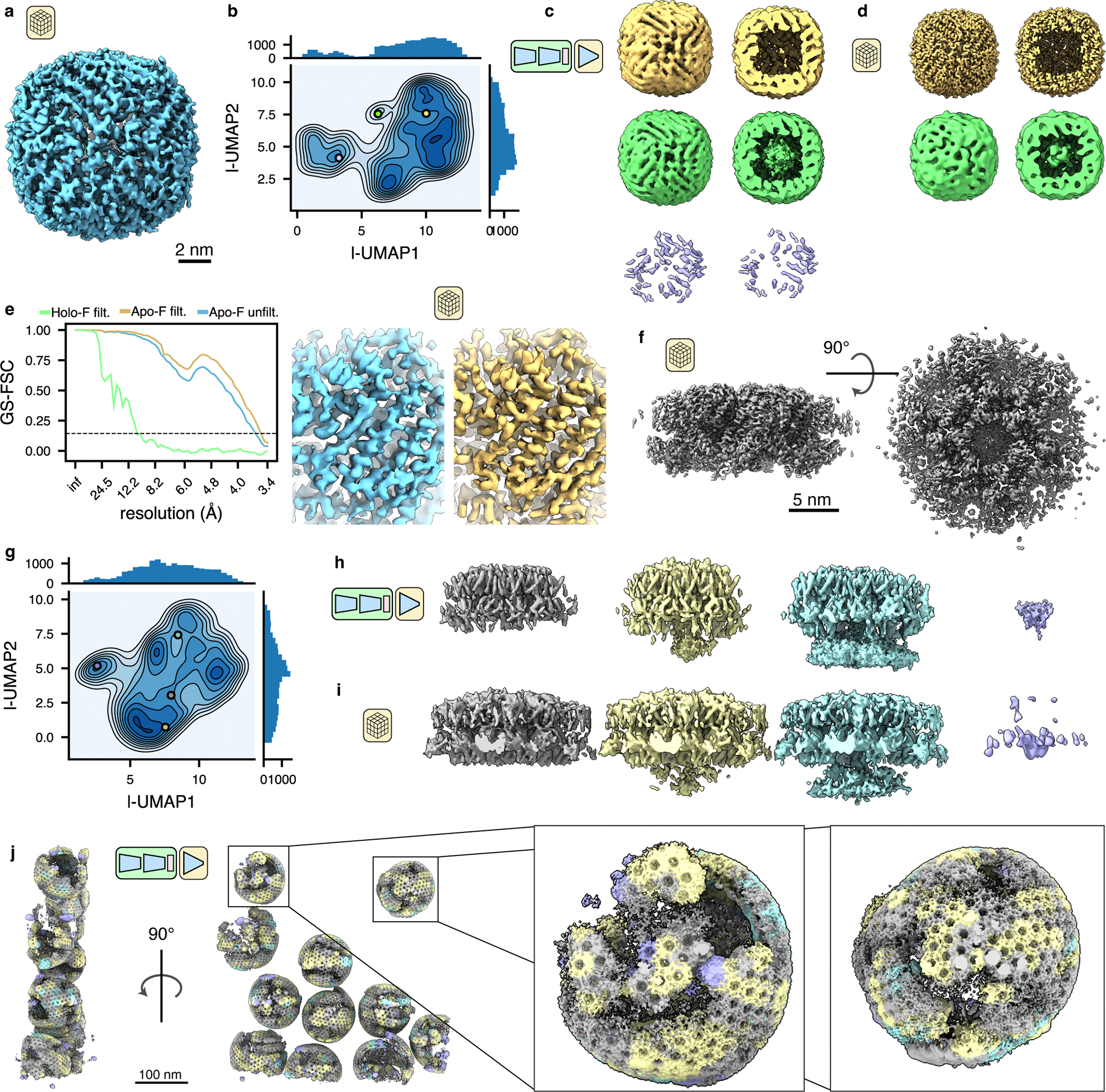
Figure 3: TomoDRGN finds residual heterogeneity within primarily-homogeneous purified particles.

(a) Consensus STA apoferritin structure refined with C1 symmetry (EMPIAR-10491, n = 25,381 particles).
(b) UMAP dimensionality reduction of tomoDRGN latent encodings from training on apoferritin dataset.
(c) Three volumes generated from tomoDRGN latent encodings sampled as indicated in (b) and rendered in their entirety (left) or clipped in plane (right).
(d) Consensus STA reconstructions of apoferritin (n = 16,576 particles; top) and iron-loaded ferritin (n = 542 particles; bottom) from multi-species refinement in M with C1 symmetry using tomoDRGN’s particle classifications, rendered at constant isosurface as in (c).
(e) Gold standard FSC curves between half-maps from the final round of M refinement with C1 symmetry for unfiltered apoferritin particles (blue) and filtered apoferritin (yellow) and iron-loaded ferritin particles (green) (left). Example of local density quality before (blue) and after (yellow) tomoDRGN particle filtering of apoferritin particles (right).
(f) Consensus STA HIV gag structure refined with C1 symmetry (EMPIAR-10164, n=18,325 particles).
(g) UMAP dimensionality reduction of tomoDRGN latent encodings from training on HIV Gag dataset.
(h) Four illustrative volumes generated from tomoDRGN latent encodings sampled as indicated in (g). Note increasing density corresponding to the lower NC layer in the yellow and cyan maps relative to that in gray.
(i) Weighted back-projection reconstructions of isolated structural classes using tomoDRGN’s particle classifications (from left to right, n = 11,449 particles, 3,546 particles, 1,444 particles, and 1,674 particle), rendered at constant isosurface.
(j) An EMPIAR-10164 tomogram reconstructed with tomoDRGN. Volumes were generated for each Gag hexamer using tomoDRGN, colored as in (h, i), and positioned correspondingly in the source tomogram. Inset highlights two representative VLPs.
