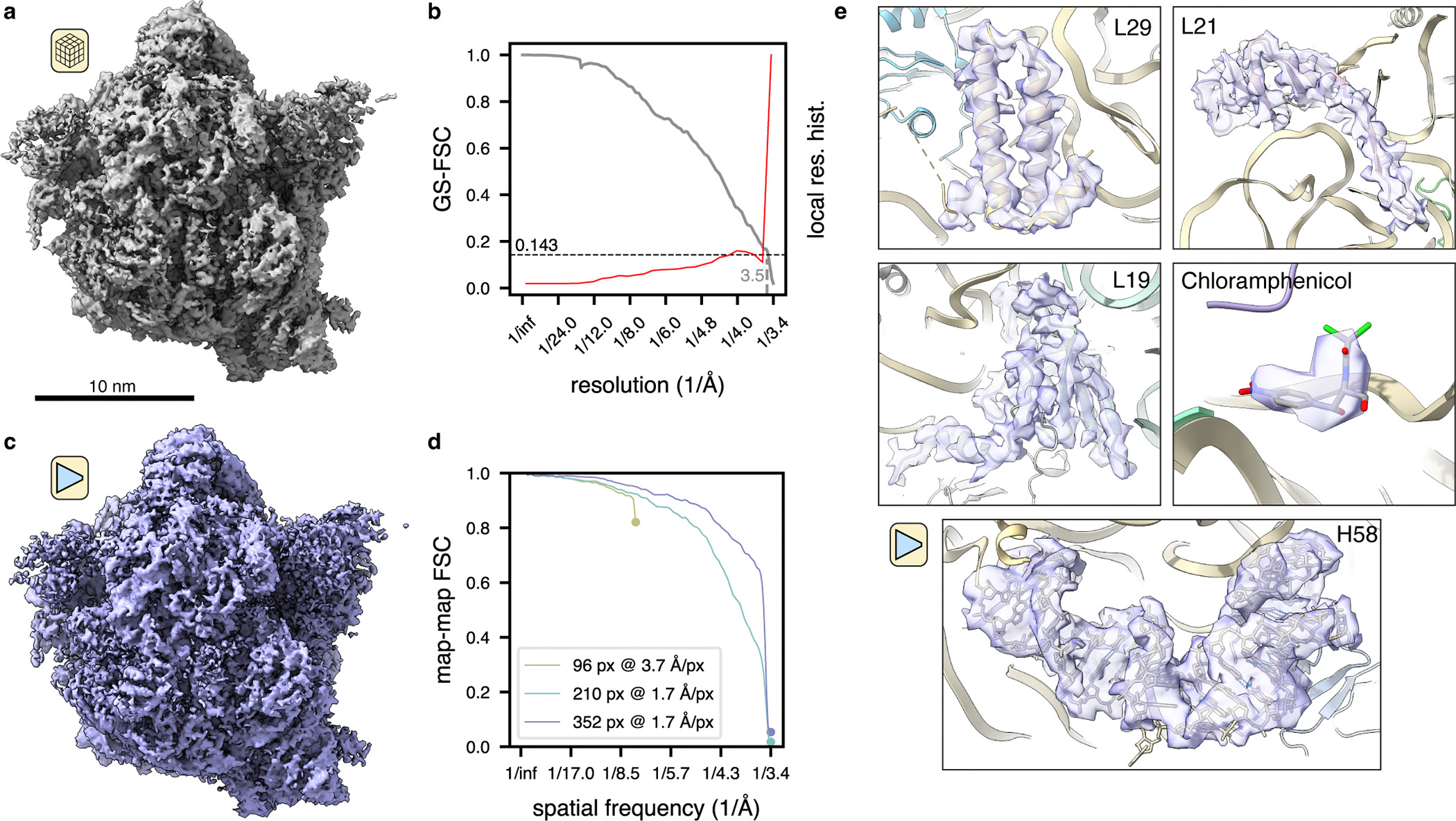
Figure 4: TomoDRGN resolves high resolution features from sub-tomograms collected in situ.

(a) M. pneumonaie ribosomal volume obtained from traditional STA processing (n=22,291 particles imaged in situ).
(b) Gold standard FSC curve between half-maps for the volume shown in (a). The second y-axis depicts a histogram of local resolution throughout the map.
(c) TomoDRGN homogeneous reconstruction of the particles used for the reconstruction in (a), lowpass filtered to 3.5Å.
(d) Map-to-map FSC of three tomoDRGN homogeneous reconstructions of the particle stack in (a) at indicated box and pixel sizes against corresponding STA volumes. Circles denote the Nyquist limit for each particle stack.
(e) Local density maps, lowpass filtered at 3.5Å, resulting from tomoDRGN homogeneous reconstruction in (c).
