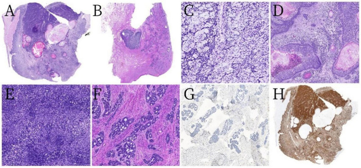
FIGURE 2.
Pathological and histological findings. (A) Tumor component one was predominantly diffuse with ill-defined margins, featuring solid sheets and nest-like arrangements in a mosaic pattern, with some areas having relatively clear boundaries (hematoxylin and eosin [H&E] staining at low magnification). (B) Tumor component two consisted of solid nodules surrounded by scattered tumor cells distributed in cribriform, tubular, solid small nest-like patterns (H&E, low magnification). (C) Mucinous stroma with a sieve-like structure (H&E, ×200 magnification). (D) Squamous epithelial differentiation with keratinization and keratin cyst formation with intraepithelial neoplasia (H&E, ×50 magnification). (E) Tumor cells were small and arranged uniformly with a high nuclear-to-cytoplasm ratio (H&E, ×200 magnification). (F) An abundance of cells in tumor component two arranged in cribriform, tubular, and solid nest-like patterns, with abundant mucin within the lumens and minimal or no stromal reaction (H&E, ×100 magnification). (G) The adenoid cribriform area was negative for CD117 by immunohistochemistry (×100 magnification). (H) All tumor cells exhibited strong diffuse positivity for p16 (low magnification).

