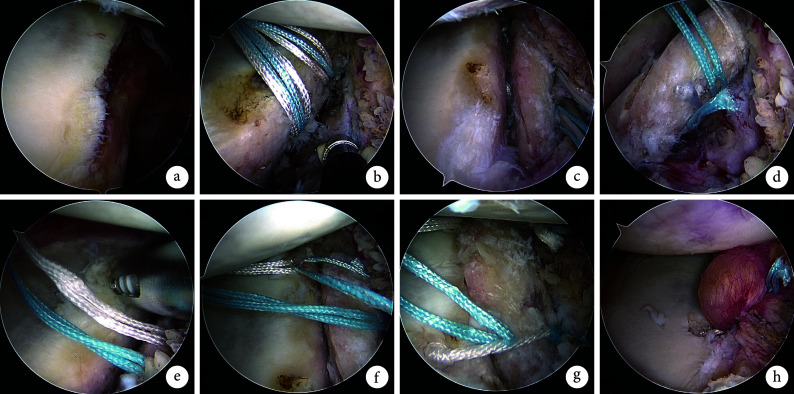
图 3.
Schematic diagram of arthroscopic bone grafting and fixation
关节镜下植骨及固定操作示意图
a. 镜下见关节盂骨缺损;b. 于骨缺损中间植入2枚盂唇钉;c. 盂唇钉缝线穿过骨块预留孔并与缺损处贴合;d. 制作第1个Pulley结以稳定骨块;e. 于关节盂骨缺损前上缘拧入1枚盂唇钉;f. 于骨块外上方制作第2个Pulley结;g. 骨块外下方制作第3个Pulley结;h. 剩余缝线进行Bankart修复
a. Arthroscopic examination revealed the glenoid bone defect; b. Two glenoid anchors were inserted in the middle of the defect; c. The suture of glenoid anchor was passed through the reserved hole and adhered to the defect; d. The first Pulley knot was made to stabilize the bone block; e. A glenoid anchor was implanted at the anterior upper edge of the glenoid defect; f. The second Pulley knot was made above the outer part of the bone block; g. The third Pulley knot was made below the outer part of the bone block; h. Bankart repair with the remaining sutures

