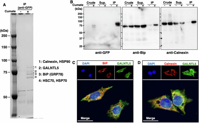
Fig. 2. The direct interaction of GALNTL5 with ER-resident chaperone proteins.
A Co-immunoprecipitants with anti-GFP antibody were separated on SDS-PAGE and visualized using the Negative Gel Stain MS Kit (Fujifilm Wako Pure Chemical Corporation). From the four fractions of visualized bands around 75 kDa, five chaperone proteins (calnexin, HSP90, BiP, HSC70, and HSP70) were identified with MS. B Anti-BiP and anti-calnexin antibodies confirmed that BiP and calnexin interact with GALNTL5. C, D Fluorescence image of GALNTL5-GFP/GC-2spd cells stained with anti-BiP antibodies, anti-calnexin antibodies, or DAPI nuclear staining, after 1 day in the presence of cumate solution. Yellow signals represent GALNTL5-GFP, and BiP or calnexin co-localization in the ER. White scale bars, 20 µm.

