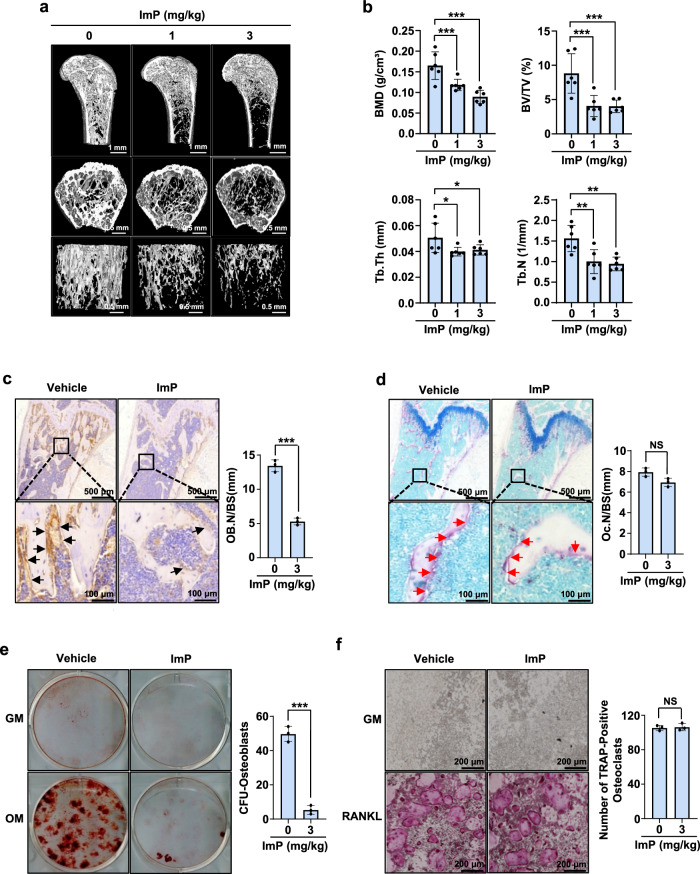
Fig. 1. ImP produces bone loss in mice.
a ImP was administered for 4 weeks using a subcutaneously implanted osmotic pump, and micro-computed tomography analysis was performed on the femurs of the mice (n = 6). b Trabecular bone parameters, including BMD (g/cm3), BV/TV (%), Tb.Th (mm), and Tb.N (1/mm) were measured using CT analyzer program (n = 6). c, d Pathological bone changes were observed through histological analysis of the samples shown in (a). Immunohistochemistry (IHC) analysis was performed using ALP staining (c) (n = 3). Additionally, TRAP staining was performed using a TRAP detection kit (d) (n = 3). e, f BMSCs and BMM cells were isolated from mice injected with ImP (3 mg/kg) for 4 weeks and identified differentiation colonies (n = 3). The results were confirmed through alizarin red S staining (e) and TRAP staining (f). ImP, imidazole propionate; BMD, bone mineral density; BV/TV, percentage bone volume; Tb.Th, trabecular thickness; Tb.N, trabecular number; IHC, Immunohistochemistry; black arrow, ALP-positive osteoblasts; red arrow, TRAP-positive osteoclasts; Values are presented as the mean ± SD; *, P < 0.05; **, P < 0.01; ***, P < 0.001; compared to the control group.

