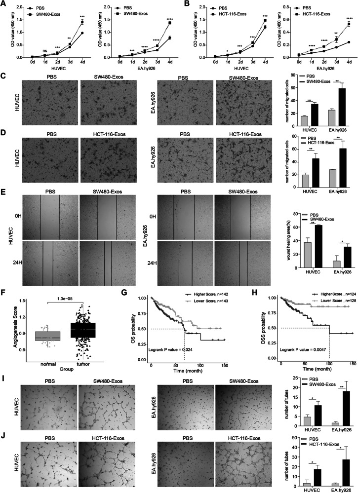
Fig. 2. Exosomes derived from colorectal cancer cells exhibit the ability to augment the proliferation, migration, and angiogenesis of vascular endothelial cells.
A, B CCK8 assay demonstrating the impact of exosomes derived from SW480 cells (A) and HCT-116 cells (B) on the proliferation of HUVEC and EA.hy926 cells. C, D Transwell assay depicting the influence of exosomes derived from SW480 cells (C) and HCT-116 cells (D) on the migration ability of HUVEC and EA.hy926 cells. E Cell wound-healing assay evaluating the effects of exosomes from SW480 on the migratory capacity of HUVEC (left) and EA.hy926 (right) cells. F Comparison of vascularity scores between paracarcinomatous and colorectal tissues. G, H Impact of high and low vasculature formation capacity scores on Overall Survival (OS) (G) and Progression-Free Survival (PFS) (H). I, J In vitro angiogenesis assay assessing the effect of exosomes from SW480 cells (I) and HCT-116 cells (J) on the angiogenic potential of HUVEC and EA.hy926 cells. (Two-tailed unpaired t-test or Mann–Whitney test. The Kaplan–Meier method was utilized to analyze survival curves, with significance evaluated using the log-rank test. *P < 0.05, **P < 0.01, ***P < 0.001, ****P < 0.0001).

