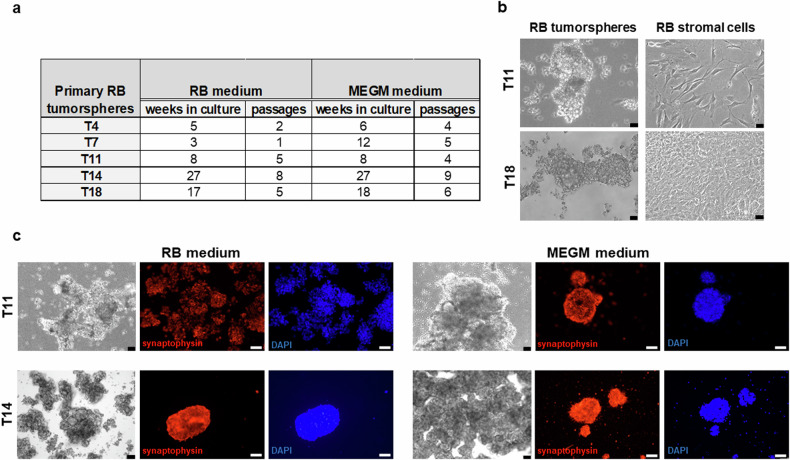
Fig. 1. Overview and comparison of primary RB tumorspheres cultured in RB and MEGM medium.
a Table summarizing weeks in culture and cell culture passages of the different RB tumorsphere specimens (T4, T7, T11, T14, T18) in the two different culture media used (RB and MEGM medium). b Phase contrast microscopy photographs comparing the morphology of unselected RB derived tumorspheres and stromal derived RB cells derived from two primary RB tumors (T11 and T18) cultured in RB medium. (scale: 64 µm). c Phase contrast imaging (black and white photos; scale bars: 64 µm) and immunofluorescence staining of two primary RB derived tumorsphere cell cultures (T11 and T14) maintained in two different growth media (RB medium in the left panel; MEGM medium in the right panel) with an antibody against synaptophysin (red fluorescence), a neuronal tumor marker and counterstained with DAPI (blue fluorescence; scale bars: 50 µm).

