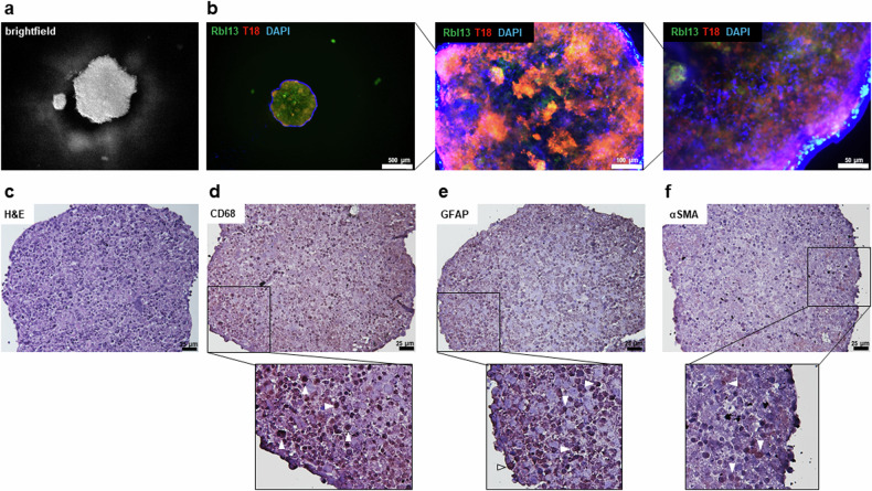
Fig. 7. Co-cultivation of RB tumor and stromal cells in a 3D cell culture model.
a Brightfield and (b) immunofluorescence pictures showing Rbl13 tumor cells in green (GFP), T18 stromal cells in red (tomato lectin) and blue nuclei counterstaining (DAPI) of a RB tumor-stroma spheroid in a hanging drop culture at different magnifications (b; scale bars: 500 µm, 100 µm and 50 µm). c Histological analysis of paraffin sections of a RB tumor-stroma spheroid stained with different indicated stromal markers and counterstained with hematoxylin and eosin (H&E; d–f; scale bars: 25 µm). White arrowheads exemplarily indicate some cells, which stained positive for the respective markers (zoomed-in insets d–f; brown signal).

