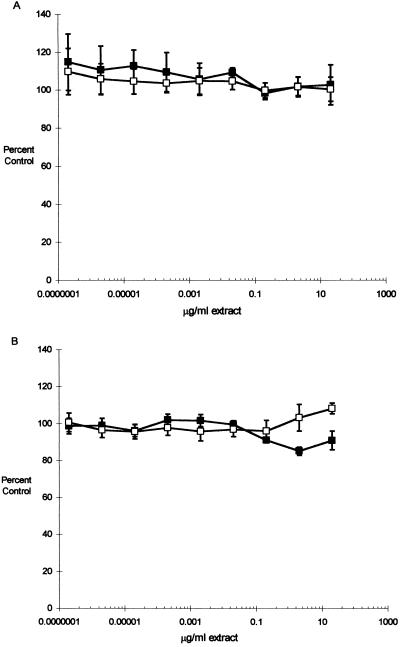
FIG. 3.
Growth and viability of bladder cells exposed to CNF1-containing (■) and control (□) extracts. T24 (A) or Hs 738 (B) cells were seeded to 96-well microtiter plates (1.0 × 10³ cells/well) and incubated with medium containing 10-fold serial dilutions of CNF1 (DH5/pISS392) or control (DH5/pGEM3) bacterial extract. At 24, 48, and 72 (shown) h, the medium in sample plates was replaced with 100 μl of fresh medium containing 20 μl of MTS solution (see the text). Incubation was continued in the dark for 4 h, and the reduction of MTS was quantitated spectrophotometrically. In each experiment, the growth in the control wells with medium containing no extract was determined and the data were expressed as percentages of control growth (means ± SEM) from two (T24 cells) or five (Hs 738 cells) experiments.

