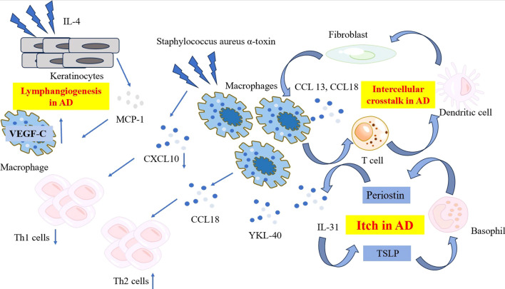
Figure 1.
The characteristic and pathogenetic roles of the macrophages in AD. Compared to psoriasis, macrophages in AD produce lower level of CXCL10 when exposed to Staphylococcus aureus α-toxin, resulting in reduced Th1 polarization. Instead, macrophages in AD produce high levels of CCL18, recruiting more Th2 cells to affected skin and release YKL-40, an important Th2 marker. A network comprising periostin, TSLP, basophils and macrophage-derived IL-31 contribute to the mechanism of itch in AD. Ligand-receptor interactions data revealed the intracellular crosstalk between CCL13, CCL18-macrohages, T cells, DCs and fibroblasts. Macrophages also get involved in the lymphangiogenesis in AD by expressing significant level of VEGF-C. AD, atopic dermatitis; CCL, C-C Motif Chemokine Ligand; CXCL, C-X-C Motif Chemokine Ligand; DC, dendritic cell; IL, interleukin; MCP-1, monocyte chemoattractant protein-1; TSLP, thymic stromal lymphopoietin; VEGF-C, vascular endothelial growth factor-C; YKL-40, Chitinase 3-like 1.

