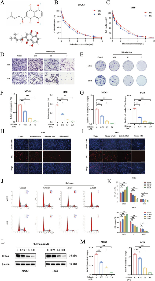
FIGURE 1.
Shikonin inhibited cell proliferation of MG63 and 143B cells. (A) The molecular and 3D structure of shikonin. (B–C) MG63 and 143B cells were cultured at different concentrations of shikonin (0, 0.25, 0.5, 1, 2, 4, 6, 8 and 10 uM) for 24, 48 h, and then CCK-8 assay was performed to detect the cell proliferation. (D) Giemsa staining was used to observe the effect of shikonin on cell morphology. (E–F) Colony formation and (G–I) EdU Staining were used to observe the impact of shikonin on cell proliferation. (J, K) Flow cytometry was used to analyze the effects of shikonin on the cell cycle when cells were treated with or without shikonin for 24 h (L, M) Western blot was used to detect the expression of proliferation protein PCNA. *p < 0.05, **p < 0.01, ***p < 0.001. The data were presented as mean ± SD, and analyzed using one-way ANOVA following Tukey’s t-test (n = 3).

