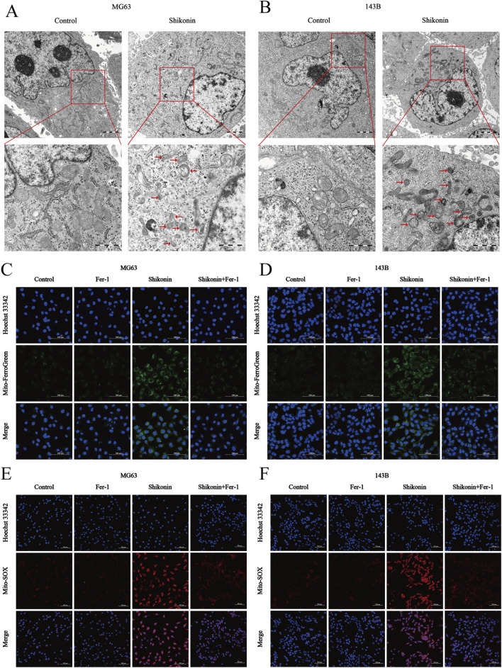
FIGURE 3.
Shikonin induced mitochondria damage in OS cells. (A, B) The mitochondrial morphology of MG63 and 143B cells treated with or without shikonin after 24 h was observed by transmission electron microscopy (scale bar = 1 um). The red arrows represented mitochondrial changes typical of ferroptosis. (C, D) Mitochondrial Fe2+ and (E, F) mitochondrial ROS contents was observed by using Laser Scanning Confocal Microscopy (LSCM) after the intervention of co-treated with shikonin and Fer-1 after 24 h in MG63 and 143B cells (scale bar = 100 um).

