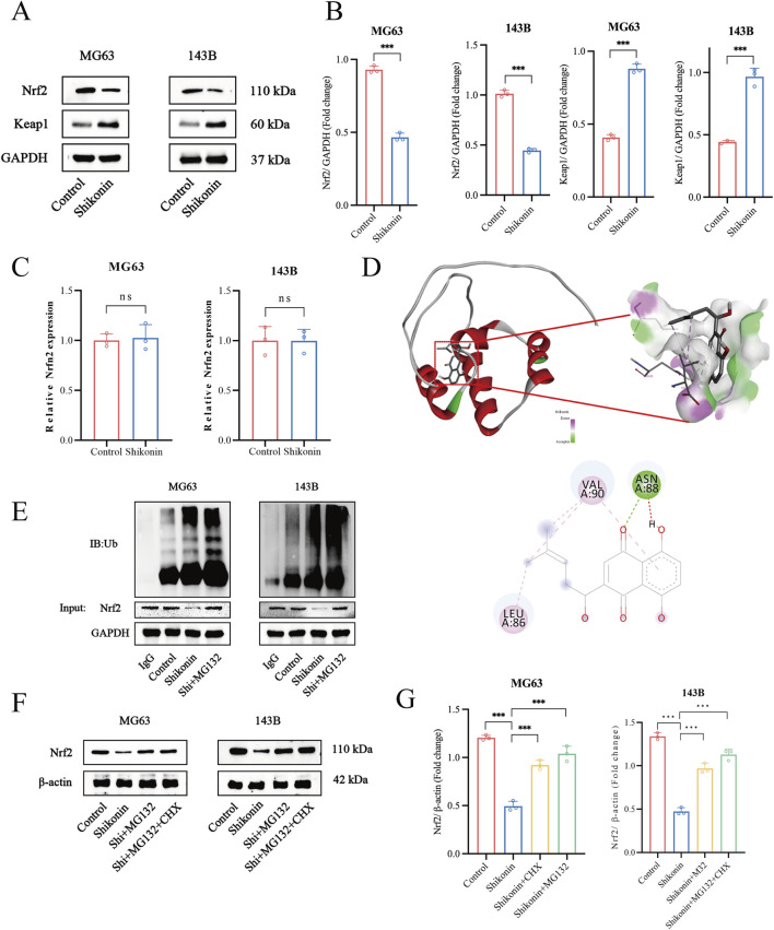
FIGURE 4.
Shikonin physically interacted with Nrf2 and promoted Nrf2 degradation in OS cells. (A, B) Western blot was used to detect the expression of Nrf2 and keap1 in shikonin-treated MG63 and 143B cells, and quantification was analyzed. (C) Nrf2 mRNA levels in the control or shikonin (1.5 uM)-treated MG63 and 143B cells for 24 h. (D) The interaction between shikonin with Nrf2 was predicted by molecular docking. (E) Co-immunoprecipitation and Western blot assay of Nrf2 protein after shikonin and MG132 (2.5 uM) co-treatment 24 h in MG63 and 143B cells. (F, G) Western blot was used to observe the expression of the Nrf2 protein after the intervention of 1.5 uM shikonin cotreated with or without MG132 (2.5 uM) and CHX (35 uM) for 24 h, and quantification was analyzed. *p < 0.05, **p < 0.01, ***p < 0.001. The data were presented as mean ± SD, and analyzed using one-way ANOVA following Tukey’s t-test (n = 3).

