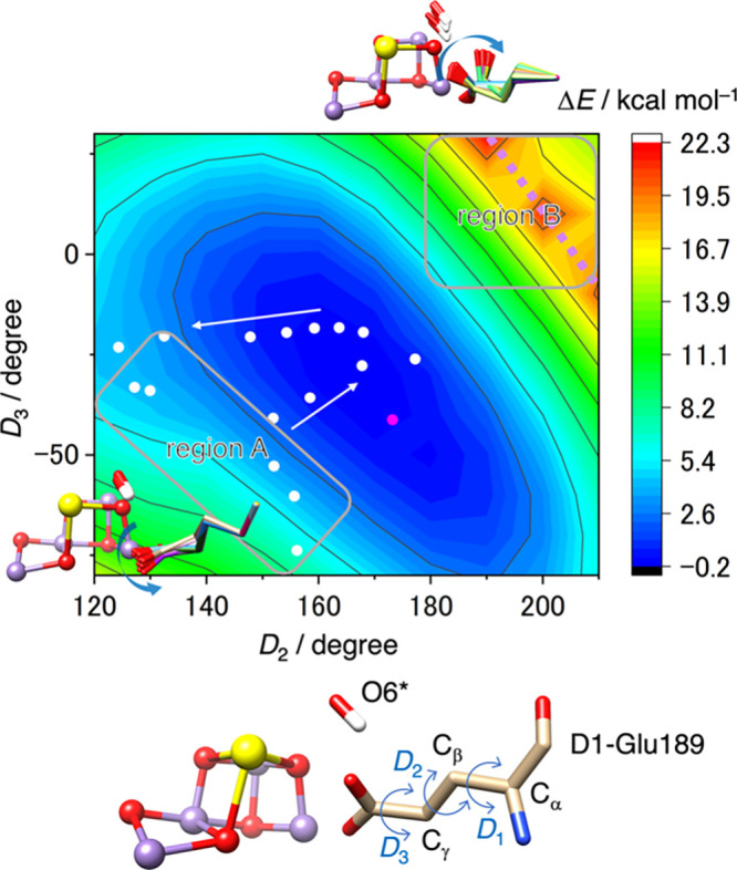
Figure 2.

A potential energy surface representing the energy change (ΔE) as a function of the dihedral angles D2 and D3 of D1-Glu189. These calculations were based on the PDB coordinates (6JLL, monomer A),10 with an additional O6* remaining attached to the Ca ion of the Mn cluster. The relative energy ΔE is referenced to the metastable structure depicted in Figure S1B. White circles indicate the D2 and D3 values corresponding to the motion of the D1-Glu189 side chain when coupled with O6* binding to MnD (see Figure 3). A magenta circle represents the D2 and D3 values observed in the S3 state, in which O5 and O6 either form an oxyl-oxo bond or exist as bridging oxo and terminal hydroxo ligands.
