Figure 3.

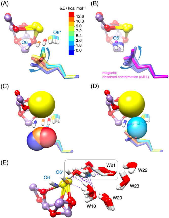
(A) Conformational changes in the side chain of D1-Glu189 as O6* moves toward MnD. The colors of O6* and D1-Glu189 correspond to the relative energy (ΔE) compared to the metastable structure shown in Figure S1B. (B) Conformational changes in the side chain of D1-Glu189 as O6* (renamed O6) binds to MnD, followed by the return of D1-Glu189 to its original orientation. The experimentally observed conformation in the S3 state (6JLL, monomer A)10 is highlighted in magenta. (C, D) A sphere representation illustrates Ca and the coordinating oxygen atoms of D1-Glu189 during O6* binding to MnD, aiding in the comprehension of the steric hindrance resulting from repulsive forces between neighboring Ca, D1-Glu189, and moving O6*. (E) Fluctuations of five water molecules (W10, W20, W21, W22, and W23) accompanying the movement of O6* toward MnD. Purple dashed lines represent hydrogen bonds between O6* and W21, as well as between O6* and W10, contributing to the stabilization of the anionic form of O6* (OH–). As O6* moves closer to MnD, W21 is also pulled toward the space between Ca and D1-Glu189 due to its interaction with O6*, while interference from D1-Glu189 (not shown) causes the hydrogen bond between O6* and W10 to break just before O6* binds to MnD (O6* → O6).
