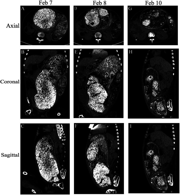
Figure 1.
Comparison of the abdominal computed tomography scan before the procedure following multiple enemas. Axial, coronal, and sagittal views were utilized for assessment. (A–C) Initial abdominal computed tomography scan revealed a discernible bowel pattern and a notable abdominal mass measuring 39.5 cm × 18.3 cm × 12.7 cm. (D–F) and (G–I) show the change in fecal characteristics after treatment.

