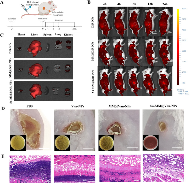
Fig. 6.
Evaluation of infection site accumulation and anti-subcutaneous infection: A. Schematic representation of experiments assessing the targeting capability of Sa-MM@Van-NPs in a mouse model of subcutaneous abscess infected by S. aureus. B. In vivo optical imaging of mice with subcutaneous abscesses following various treatments at 2, 4, 8, 12, and 24 h post-injection. C. Ex vivo imaging of DiR-labeled nanoparticles in major organs at 8 h post-injection. D. Representative images of skin lesions and bacterial colonies (lower left corner) in infected tissues under different treatments on day 4 (scale bar = 5 mm). E. H&E staining of skin tissue sections on day 4 (scale bar = 100 μm)

