
FIGURE 1.
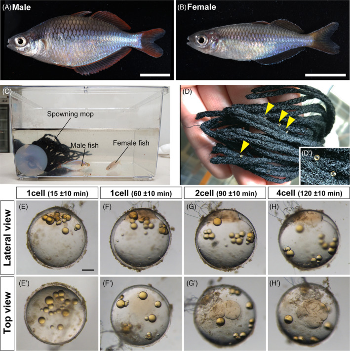
Collection of fertilized eggs in Melanotaenia praecox. (A), (B) Representative images of adult males (A) and females (B). (C) Each mature male and female pair was kept in a 3 L breeding tank containing a spawning mop. (D), (D’) Eggs attached to the spawning mop by their attaching filaments. A magnified view of the attached eggs (D’). Yellow arrowheads indicate the attached eggs. (E)–(H’) Progress in the first and second cleavages of an M. praecox egg at the one‐cell stage, 15 ± 10 (E), (E’) and 60 ± 10 min (F), (F’) after fertilization; the two‐cell stage at 90 ± 10 min (G), (G’); and the four‐cell stage at 120 ± 10 min (H), (H’). Scale bar indicates 1 cm (A), (B) and 200 μm (E).