FIGURE 2.

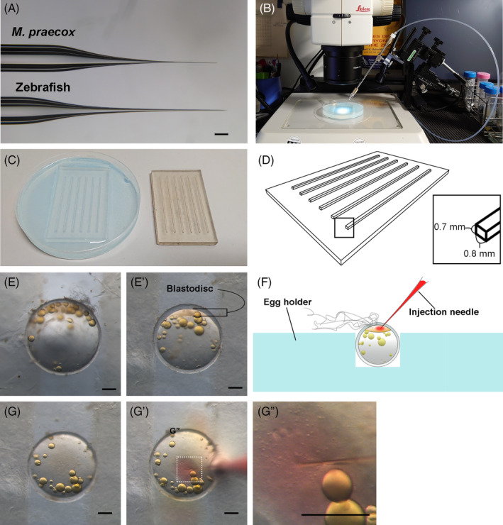
Microinjection of fertilized eggs. (A) A comparison of a microinjection needle for Melanotaeni praecox (top) with that for zebrafish (bottom). (B) Microinjection set‐up. Needle holder set‐up with a 3D micromanipulator to precisely control the position of the needle under a stereomicroscope. (C) A 3D‐printed mold (right) and an egg holder made of 3% agarose (left). (D) Protrusions on the 3D‐printed mold were 0.8 mm in width and 0.7 mm in height. (E), (E’) A comparison of the diameter of an M. praecox egg with the width of a groove in the egg holder. Images of an egg not held (E) and held (E’) in the groove. (F) A schematic illustration of the microinjection. (G)–(G”) Successful injection through the chorion to the cytoplasm was visualized by red coloration of phenol red contained in the injection solution. Scale bars indicate 200 μm.
