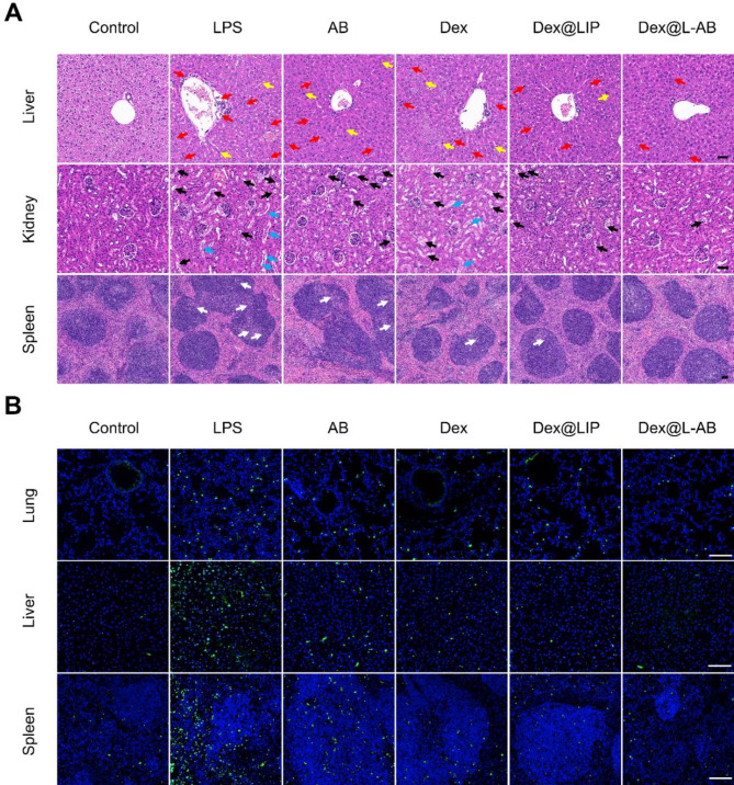
Fig. 7.
Dex@L-AB relieved tissue injuries and cellular apoptosis of septic mice. (A) H&E staining of liver, kidney and spleen tissues. Red arrows indicate inflammatory cells infiltration; yellow arrows indicate liver necrosis cells; black arrows indicate vacuolated renal tubules; blue arrows indicate necrotic shedding of renal tubular epithelial cells to form casts and white arrows indicate disorganized germinal centers. (B) TUNEL staining of lung, liver and spleen tissues (green fluorescence: TUNEL positive cells; blue fluorescence: cell nucleus). Scale bar = 100 μm

