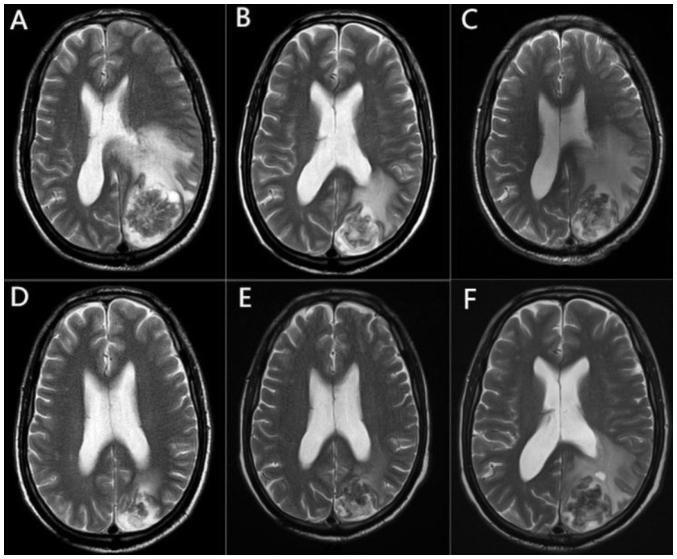
Figure 1.
Brain MRI changes in the patient throughout the course of the disease. (A) MRI presentation at the earliest detection of brain metastases. (B) MRI after radiotherapy treatment of the brain lesion, showing reduced metastasis and surrounding edema. (C) MRI at the time of brain radiation necrosis diagnosis, showing a significant increase in edema around the lesion. (D) MRI after two cycles of bevacizumab treatment, showing a decrease in edema around the lesion. (E) MRI after four cycles of bevacizumab treatment, with a slight decrease in the perifocal edema and a significant reduction in edema compared to before bevacizumab treatment. (F) MRI at diagnosis of recurrent brain radiation necrosis, showing an increase in edema around the intracranial lesion. MRI, magnetic resonance imaging.

