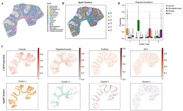
Fig. 5.
The results of Spall for mouse cerebellum. A Pie plots showing the cell type composition at each spot, deconvolved by Spall from cerebellar tissue data. B Spatial domain identification based on cell type proportions estimated by Spall. C Top: spatial proportions of four layer-specific cell types predicted by Spall. Bottom: Corresponding spatial domains identified using proportions predicted by Spall. D Cell type abundance analysis across the four spatial domains. The center line represents the median, box limits indicate the upper and lower quartiles, and whiskers correspond to 1.5 × the interquartile range

