Fig. 1.
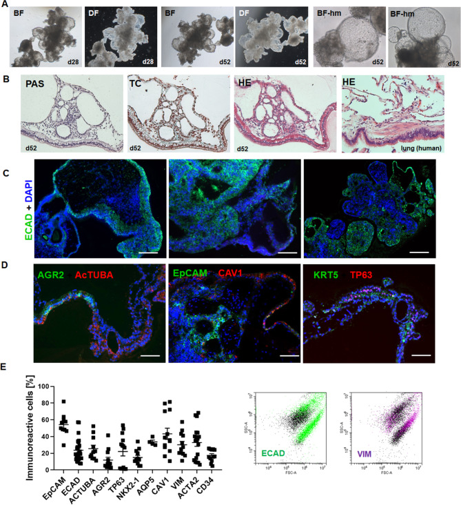
Morphological characterization of free-floating iPSC-derived human LuOrgs confirmed the efficient generation of desired lung structures and cell types. (A) Bright-field (BF) and dark-field (DF) images of LuOrgs generated matrix-free from human iPSCs in ultra-low attachment plates at indicated time points using the 5x and 20x objective of an inverted microscope. (B) Histology staining of paraffin-embedded sections of the lung-like regions of LuOrgs using Periodic Acid Schiff (PAS), trichrome (TC), and hematoxylin and eosin (HE) staining. Normal human lung tissue was included as positive control. Magnification: 10x. (C) Immunofluorescence staining of paraffin-embedded sections of LuOrgs representing different the lung-like regions for the lung epithelial markers ECAD (green). Nuclei were counterstained using DAPI (blue). Scale bar: 200 μm (D) Representative immunofluorescent images of indicated lung cell type-marker proteins. Scale bar: 100 μm. (E) Flow cytometry quantification of epithelial and mesodermal lung cell types of single cell dissociated LuOrgs (d45-52). Data are shown as means ± SEM of indicated biological replicates. Representative FACS plots of an epithelial and mesodermal marker is shown. See also Figure S1

