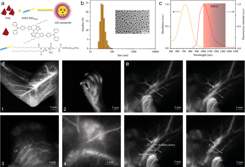
Fig. 11.
a The preparation of AIE probes via a nanoprecipitation method. b Dynamic light scattering data of aqueous AIE probes. Inset: transmission electron microscopy image of water-dispersed AIE probes. c Fluorescence and absorption spectra of AIE probes. d NIR-II fluorescence imaging of the vessels and lymph nodes in cynomolgus monkey after intravenous injection of the AIE probes. (1) arm, (2) hand, (3) scalp vasculature, (4) an axillary lymph node marked by the red dotted line. e NIR-II fluorescence imaging of the deep axillary artery in a healthy adult cynomolgus monkey after intravenous injection of AIE probes. When epidermis was moved, the superficial vein was moved accordingly; however, the deep axillary artery remained stationary. Reproduced with permission from Tang et al. [121]

