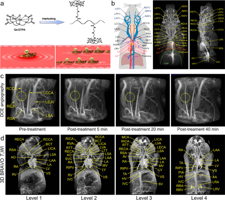
Fig. 2.
a The schematic illustration of zwitterionic polymeric MR contrast agents PAA-Gd compared with individual Gd-DTPA. b Schematic drawing of vascular anatomy and the vascular identification in different directions of 3D angiography of mouse body. c PAA-Gd enhanced 3D angiography of mouse carotid thrombosis pre-treatment (obtained at 5 min after PAA-Gd injection), and the real-time monitoring of thrombolytic therapy post-treatment of urokinase‐type plasminogen activator (uPA). (The carotid thrombosis was identified by yellow dotted circle). d Different coronal levels of 3D BRAVO images of swine for identifying the vascular structures. Reproduced with permission from Hou et al. [45]

