
FIGURE 5.
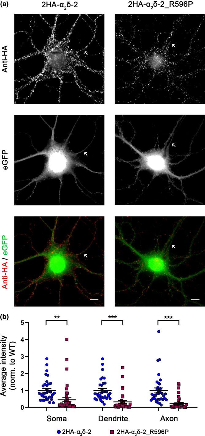
Strongly reduced membrane expression of α2δ‐2_R596P in differentiated cultured hippocampal neurons. (a) Representative examples of primary cultured hippocampal neurons transfected with soluble eGFP together with either HA‐tagged WT (2HA‐α2δ‐2) or mutated (2HA‐α2δ‐2_R596P) α2δ‐2. Anti‐HA live‐cell labeling demonstrates a reduced staining intensity of α2δ‐2_R596P in the soma, dendrites, and the axon (indicated by an arrow) compared to WT α2δ‐2. (b) Quantification of the average HA fluorescent intensities in the three compartments shows that the surface expression of mutated α2δ‐2 is strongly reduced compared to WT α2δ‐2. Graphs show values for individual cells (dots) and mean ± SEM (lines). All values were normalized to the mean of the WT 2HA‐α2δ‐2 fluorescence intensity within each culture preparation. Data were obtained from three independent culture preparations; 35 and 45 cells expressing WT or mutated HA‐tagged α2δ‐2 were analyzed, respectively. Statistics: Unpaired t‐test, soma: T (78) = 3.4; **p = 0.0011, dendrite: T (78) = 5.1; ***p < 0.0001, axon: T (78) = 5.5; ***p < 0.0001. Scale bars, 10 μm.