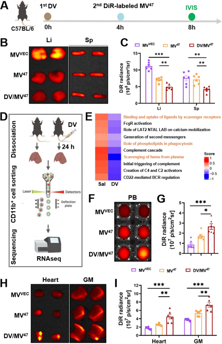
Fig. 4.
In vivo biodistribution of therapeutic MV47. (A) Schematic illustration of in vivo testing: C57BL/6 mice received tail vein injections of DiR-labelled MVVEC and MV47, with or without DV pre-blocking, for subsequent in vivo tracking. (B&C) Representative DiR optical images alongside quantification of biodistribution in the liver and spleen (n = 6). (D) Schematic detailing the sample preparation process for RNAseq. (E) Gene set variation analysis (GSVA) was conducted on macrophage activity-related Reactome pathways, highlighting phagocytosis-related pathways in orange. (F-I) Representative DiR optical images and accompanying quantification for biodistribution in the peripheral blood (F&G), heart, and gastrocnemius muscle (H&I) (n = 6). Li, liver; Sp, spleen; PB, peripheral blood; GM, gastrocnemius muscle. Statistical evaluations were conducted using one-way ANOVA. Results were presented as mean ± SEM (* P < 0.05; ** P < 0.01; *** P < 0.001)

