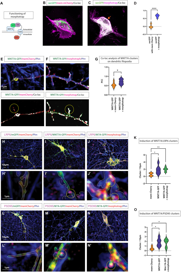
Fig. 3.
Membrane-tethered WNT7A-GFP can cluster Wnt and synaptic components. (A) Schematic of the tethering experiment using double transfection of WNT7A-GFP and the morphotrap nanobody construct, variable heavy domain of the heavy chain (Vhh)-CD8-mCherry, which binds GFP-tagged proteins. (B,C) AGS cells transfected with soluble-GFP (secGFP) and mem-mCherry (B) and AGS cells transfected with secGFP and morphotrap (C). Orange arrow indicates membrane localisation of secGFP. (D) Quantification identifies a significant colocalisation between the normally secreted secGFP and the morphotrap at the plasma membrane. An unpaired, one-tailed Student's t-test (****P<0.001). (E) The colocalisation of mem-mCherry and WNT7A-GFP on protrusions is shown in white. (F,G) Significantly higher WNT7A-GFP colocalisation on filopodia is observed when co-transfected with morphotrap (F), quantified by Pearson's correlation coefficient (PCC; G). Unpaired, one-tailed Student's t-test (*P<0.05). (H,H′) Super-resolution imaging of iPSC-derived cortical neurons transfected with mem-GFP and mem-mCherry, followed by post-staining for LRP6 and the actin cytoskeleton (phalloidin, Phn). (I,I′) Transfection with WNT7A-GFP and mem-mCherry, followed by post-staining for LRP6 and the actin cytoskeleton identified WNT7A-GFP-positive protrusions that cluster LRP6 in apposed cells (I′, yellow circle). (J,J′) Transfection with WNT7A-GFP and morphotrap, followed by post-staining for LRP6 and cytoskeleton, identified protrusions harbouring membrane-tethered WNT7A-GFP that cluster LRP6 in apposed cells (J′, yellow circles). (K) Quantification of co-clustering of LRP6 with WNT7A-GFP identified a significant increase in both the morphotrap and mem-mCherry conditions compared to mem-Ch alone. Unpaired, one-tailed Student's t-test (*P<0.05, **P<0.01). (L,L′) Transfection with mem-GFP and mem-mCherry, followed by post-staining for PSD95 and the actin cytoskeleton (Phn). (M,M′) Transfection with WNT7A-GFP and mem-mCherry, followed by post-staining for PSD95 and actin, revealed colocalisation on dendritic protrusions (M′, yellow circle). (N,N′) WNT7A-GFP co-transfected with morphotrap and colocalised with PSD95 on dendritic protrusions (N′, yellow circle). (O) Quantification of the colocalisation of PSD95 with WNT7A-GFP also identified a significant increase in both the morphotrap and mem-mCherry conditions compared to mem-Ch alone. Experiments were performed on three independent biological replicates. A minimum of three transfected neurons were quantified per condition. Statistical significance was addressed using one-way ANOVA with Dunnett's post-hoc test for multiple comparisons, comparing groups to the mem-mCherry/mem-GFP control group. *P<0.05. In H-J,L-N, boxed areas indicate the area shown at higher magnification in H′-J′,L′-N′.

