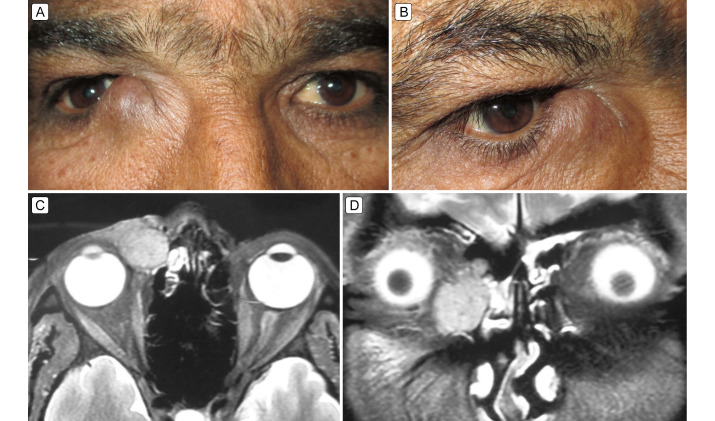
Figure 1.
A, The right lacrimal sac region shows an oval, grape-sized mass (25 × 21 mm) extending above the medial canthal tendon, pushing it superiorly; the right globe shows mild lateral displacement. B, Oblique view showing overlying skin with a bluish appearance, stretched, and free from mass. C, Contrast-enhanced magnetic resonance imaging (axial section) of the orbits showing a well-defined hyperintense mass in the lacrimal sac fossa, with mild lateral globe dystopia; there is regional ethmoid sinus hyperintensity. D, Coronal section showing a mass in the lacrimal sac region, with free nasolacrimal duct. The surrounding ethmoid sinus haze is indicated.

