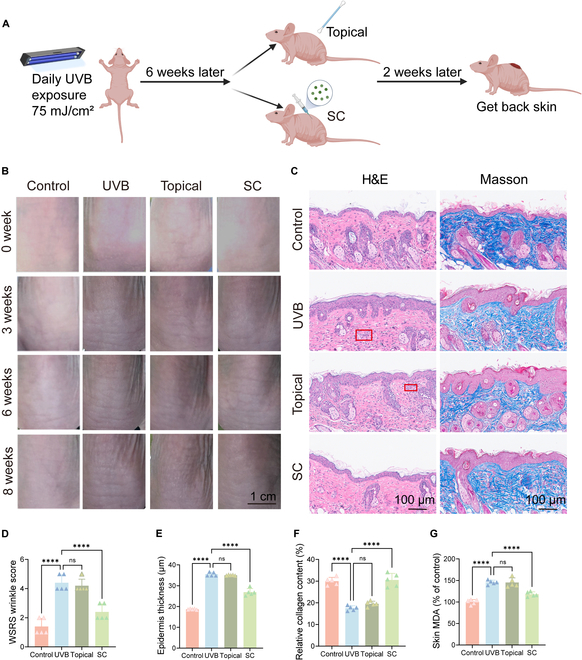
Fig. 4.

Effects of PMELNVs on the nude mouse photoaging model. (A) Schematic diagram of photoaging modeling in mice. (B) Changes in the dorsal skin of nude mice. (C) H&E and Masson staining. (D) Skin wrinkle scores. (E and F) Quantitative analysis of epidermal thickness using H&E staining and collagen deposition using Masson staining. (G) Skin MDA levels.
